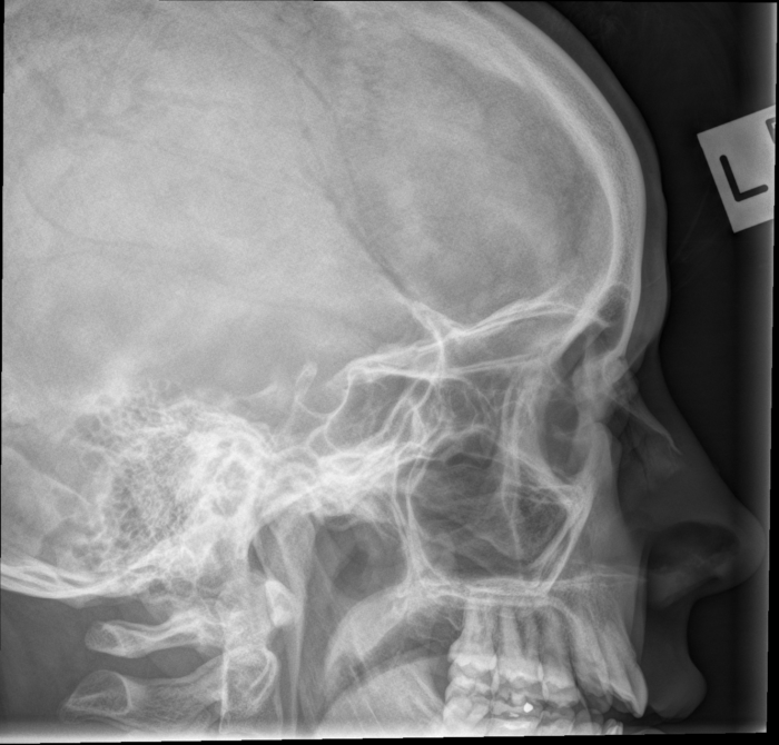
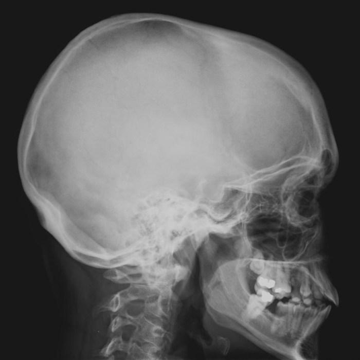
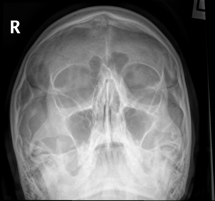
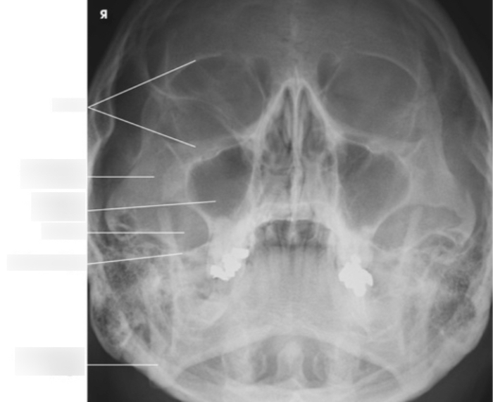
Lateral facial bones x ray – Embark on an intriguing journey into the realm of lateral facial bones x-ray, where we uncover the anatomical intricacies of these remarkable structures. From the maxillary bone’s intricate processes and foramina to the zygomatic bone’s distinctive shape, this narrative delves into the depths of their structure and function.
Our exploration continues with an examination of the diverse imaging techniques employed to visualize these bones, each with its own advantages and limitations. We delve into the optimal parameters for lateral facial bone evaluation, ensuring precise and informative imaging.
Lateral Facial Bones
The lateral facial bones form the sides of the face and include the maxillary bone and the zygomatic bone. These bones provide support for the facial muscles and protect the underlying structures.
Maxillary Bone
The maxillary bone is the largest bone of the face. It forms the upper jaw and the floor of the orbit. The maxillary bone has several processes and foramina, including:
- Frontal process:Extends medially to form part of the nasal cavity.
- Zygomatic process:Projects laterally to articulate with the zygomatic bone.
- Alveolar process:Bears the teeth of the upper jaw.
- Palatine process:Forms the hard palate.
- Infraorbital foramen:Transmits the infraorbital nerve and vessels.
- Maxillary sinus:A large air-filled cavity within the bone.
Zygomatic Bone
The zygomatic bone is a small, triangular bone that forms the prominence of the cheek. It articulates with the maxillary bone, the frontal bone, and the temporal bone. The zygomatic bone provides attachment for the muscles of facial expression.
Imaging Techniques for Lateral Facial Bones: Lateral Facial Bones X Ray

Lateral facial bones are visualized using various imaging techniques. Each technique offers unique advantages and limitations, affecting the choice of imaging modality for specific clinical scenarios.
Radiography
- Advantages: Widely available, low cost, portable, provides quick results.
- Limitations: Limited soft tissue visualization, superimposition of structures, poor visualization of complex anatomy.
Computed Tomography (CT)
- Advantages: Excellent bone visualization, allows multiplanar reconstructions, provides detailed anatomical information.
- Limitations: Higher radiation dose, potential for artifacts, longer scan time.
Magnetic Resonance Imaging (MRI)
- Advantages: Excellent soft tissue visualization, allows differentiation between normal and pathological tissues.
- Limitations: Lower bone visualization, higher cost, longer scan time, contraindicated in patients with metal implants.
Optimal Imaging Parameters
Optimal imaging parameters for lateral facial bone evaluation include:
- Collimator: Use a collimator to minimize scatter radiation and improve image quality.
- Exposure factors: Adjust exposure factors (kVp, mAs) to optimize bone visualization and minimize patient dose.
- Positioning: Position the patient correctly to minimize superimposition and ensure adequate visualization of the region of interest.
Radiographic Anatomy of Lateral Facial Bones

Radiography plays a crucial role in assessing the lateral facial bones, providing valuable insights into their anatomical structures and any potential abnormalities.
The maxillary and zygomatic bones, in particular, are commonly visualized on lateral facial bone X-rays. Understanding their radiographic features is essential for accurate interpretation.
Radiographic Comparison of Maxillary and Zygomatic Bones
The following table summarizes the key radiographic features of the maxillary and zygomatic bones:
| Feature | Maxillary Bone | Zygomatic Bone |
|---|---|---|
| Location | Forms the upper jaw and part of the orbital floor | Forms the prominence of the cheek and part of the lateral orbital wall |
| Shape | Triangular, with a thick body and alveolar process | Quadrilateral, with a thin body and zygomatic arch |
| Margins | Smooth superior and inferior margins; serrated nasal margin | Sharp lateral margin; smooth medial and inferior margins |
| Internal Structures | Maxillary sinus, nasal cavity, and alveolar sockets | None |
Pathologies of Lateral Facial Bones

The lateral facial bones are susceptible to a range of pathological conditions that can affect their structure and function. These conditions can arise from trauma, infection, or neoplastic processes, and their radiographic manifestations provide valuable insights for diagnosis and monitoring.
Common pathologies of the lateral facial bones include:
- Fractures
- Infections
- Tumors
Fractures
Fractures of the lateral facial bones are commonly caused by trauma, such as falls, sports injuries, or assaults. Radiographically, fractures appear as linear or comminuted breaks in the bone structure, with varying degrees of displacement. The location and severity of the fracture can influence the treatment plan and prognosis.
Infections
Infections of the lateral facial bones, known as osteomyelitis, can be caused by bacterial or fungal organisms. Radiographically, osteomyelitis typically manifests as areas of bone destruction or lucency, with surrounding sclerosis or thickening. The presence of soft tissue swelling or abscess formation may also be evident.
When examining lateral facial bones x-ray, it’s important to understand the role of Robert Walton Apex, an anatomical landmark that helps radiologists accurately assess facial structures. For more information on Robert Walton Apex, please refer to this article . By understanding this landmark, radiologists can provide precise interpretations of lateral facial bones x-rays, aiding in the diagnosis and treatment of facial injuries and conditions.
Tumors
Tumors of the lateral facial bones can be either benign or malignant. Benign tumors, such as osteomas or fibrous dysplasia, appear as well-defined lesions with slow growth and minimal surrounding bone destruction. Malignant tumors, such as osteosarcoma or chondrosarcoma, exhibit more aggressive radiographic features, including rapid growth, bone destruction, and soft tissue involvement.
Clinical Applications of Lateral Facial Bone Imaging

Lateral facial bone imaging plays a crucial role in diagnosing and managing a wide range of clinical conditions. It provides valuable insights into the anatomy and pathology of the facial bones, aiding in treatment planning and monitoring.
Diagnosis of Facial Fractures
Lateral facial bone radiographs are essential for detecting and assessing facial fractures. The clear visualization of bony structures allows clinicians to identify the location, extent, and severity of fractures, guiding appropriate treatment decisions. Accurate imaging interpretation is crucial to determine whether conservative management or surgical intervention is necessary.
Evaluation of Facial Trauma, Lateral facial bones x ray
In cases of facial trauma, lateral facial bone imaging helps evaluate the extent of injuries and assess the involvement of underlying structures. It assists in identifying soft tissue swelling, hemorrhage, or foreign bodies, providing a comprehensive picture of the trauma’s impact.
Assessment of Dental Impactions
Lateral facial bone imaging is vital in diagnosing and managing impacted teeth, especially those in the maxilla and mandible. The images reveal the position and orientation of the impacted tooth, aiding in treatment planning and surgical extraction procedures.
Evaluation of Facial Asymmetry
Lateral facial bone radiographs can help determine the cause of facial asymmetry, whether due to developmental anomalies, trauma, or other pathological conditions. The images provide a detailed view of the facial bones, allowing clinicians to assess symmetry and identify any abnormalities.
Monitoring of Treatment Outcomes
Lateral facial bone imaging is used to monitor the progress of treatment for various facial conditions. It helps evaluate the healing process after fractures, assess the effectiveness of orthodontic interventions, and track the results of surgical procedures, ensuring optimal patient outcomes.
General Inquiries
What are the common pathologies that affect lateral facial bones?
Common pathologies include fractures, infections, and tumors, each with distinct radiographic manifestations.
How can imaging be used to diagnose and monitor these conditions?
Imaging provides valuable insights into the extent and severity of pathologies, aiding in diagnosis and guiding treatment decisions.
What is the importance of accurate interpretation and reporting of lateral facial bone radiographs?
Precise interpretation and reporting are crucial for proper diagnosis and appropriate patient management.