Bone Markings of the Skull Quiz: Delve into the fascinating world of craniology, where you’ll unravel the secrets hidden within the intricate bone markings of the skull. Discover the significance of these anatomical features and embark on a journey of exploration that will leave you with a deeper appreciation for the human body.
From the interlocking sutures that connect skull bones to the foramina and canals that provide passage for nerves and blood vessels, this quiz will guide you through the diverse array of bone markings found on the human skull. Get ready to test your knowledge and expand your understanding of this captivating subject.
Skull Bone Markings Overview

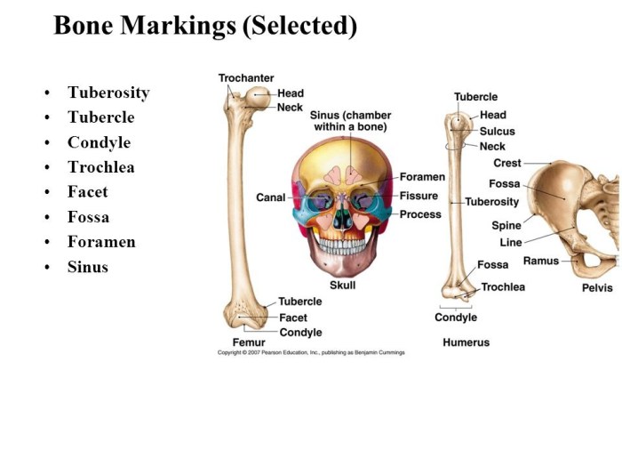
Bone markings on the skull are significant as they serve as attachment points for muscles, ligaments, and tendons, and provide passageways for nerves and blood vessels. These markings facilitate the skull’s structural integrity, functionality, and interaction with surrounding tissues.Bone markings are anatomical features found on the surface of bones, including the skull.
They encompass a wide range of structures, including depressions, foramina, processes, and sutures. These markings vary in size, shape, and location, each serving a specific purpose.
Types of Bone Markings
- Depressions: These are indentations or concavities on the bone surface, providing attachment sites for muscles and ligaments. Examples include the temporal fossa and mandibular fossa.
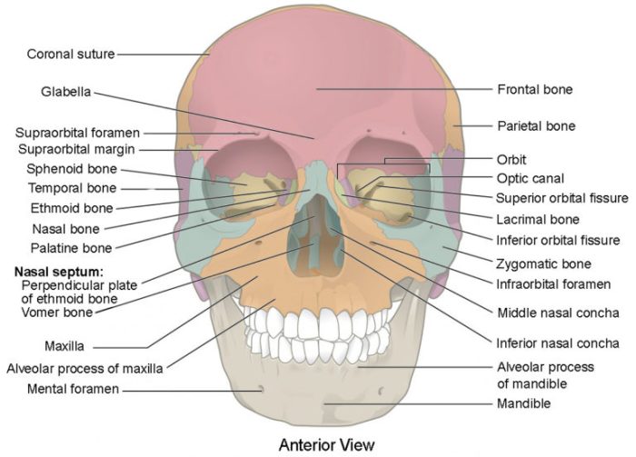
- Foramina: These are openings or holes in the bone, allowing nerves and blood vessels to pass through. Examples include the foramen magnum and supraorbital foramen.
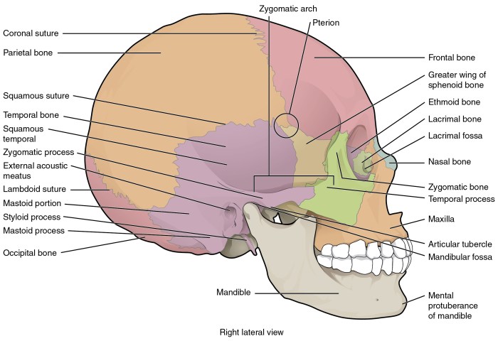
- Processes: These are projections or outgrowths of bone, serving as attachment points for muscles and tendons. Examples include the mastoid process and zygomatic process.
- Sutures: These are immovable joints between skull bones, formed by interlocking edges. Examples include the coronal suture and sagittal suture.
Sutures and Joints
Sutures are immovable joints that connect the bones of the skull. They allow for some movement and growth of the skull, but they also provide strength and stability.
There are three main types of sutures on the skull: coronal, sagittal, and lambdoid. The coronal suture runs across the top of the skull, connecting the frontal bone to the parietal bones. The sagittal suture runs down the middle of the skull, connecting the two parietal bones.
The lambdoid suture runs across the back of the skull, connecting the parietal bones to the occipital bone.
Cranial Joints
In addition to sutures, the skull also has two movable joints: the temporomandibular joint (TMJ) and the atlanto-occipital joint. The TMJ connects the mandible (lower jaw) to the temporal bone. It allows for the movement of the jaw, which is necessary for speaking, eating, and yawning.
The atlanto-occipital joint connects the skull to the first cervical vertebra (C1). It allows for the nodding movement of the head.
Foramina and Canals

The skull features numerous foramina (holes) and canals (passages) that allow for the passage of nerves and blood vessels. These structures play a crucial role in the functioning of the brain and other structures within the skull.
The major foramina and canals of the skull include:
Foramina
- Foramen magnum:Located at the base of the skull, it allows the passage of the spinal cord.
- Foramen ovale:Located on the sphenoid bone, it allows the passage of the mandibular nerve and accessory meningeal artery.
- Foramen rotundum:Also located on the sphenoid bone, it allows the passage of the maxillary nerve.
- Foramen lacerum:Located between the sphenoid and temporal bones, it allows the passage of the internal carotid artery and nerves.
- Optic foramen:Located on the sphenoid bone, it allows the passage of the optic nerve.
- Jugular foramen:Located on the occipital bone, it allows the passage of the jugular vein and nerves.
Canals
- Carotid canal:Located in the temporal bone, it transmits the internal carotid artery.
- Hypoglossal canal:Located in the occipital bone, it transmits the hypoglossal nerve.
- Condylar canal:Located in the occipital bone, it transmits the condylar nerve.
- Mastoid canal:Located in the temporal bone, it transmits the facial nerve and stylomastoid artery.
- Auditory canal:Located in the temporal bone, it transmits sound waves to the inner ear.
Processes and Projections: Bone Markings Of The Skull Quiz

Processes and projections are bony outgrowths that extend from the surface of the skull. They serve various functions, including providing attachment points for muscles, ligaments, and tendons; protecting underlying structures; and providing passages for nerves and blood vessels.
Types of Processes and Projections
- Crista: A sharp, narrow ridge, e.g., the supraorbital crest on the frontal bone.
- Tubercle: A small, rounded elevation, e.g., the mastoid tubercle on the temporal bone.
- Spine: A sharp, pointed projection, e.g., the nasal spine on the maxilla.
- Process: A general term for any bony outgrowth, e.g., the zygomatic process of the temporal bone.
Functions and Locations
Processes and projections are found throughout the skull, serving specific functions depending on their location. For instance, the styloid process of the temporal bone provides an attachment point for muscles involved in swallowing and tongue movement. The coronoid process of the mandible forms the anterior boundary of the mandibular notch and provides an attachment point for the temporalis muscle.
Examples
* Supraorbital crest: Protects the eyes and provides an attachment point for the frontalis muscle.
Mastoid process
Provides an attachment point for muscles involved in head movement and supports the ear canal.
Nasal spine
Forms the anterior end of the nasal septum and provides an attachment point for the cartilaginous part of the nose.
Styloid process
Supports the tongue and provides an attachment point for muscles involved in swallowing.
Coronoid process
Assists in closing the jaw and provides an attachment point for the temporalis muscle.
Depressions and Grooves

Depressions and grooves are indentations on the skull that serve various functions, including muscle attachment and nerve passage. These features provide structural support and facilitate movement and sensation in the head and neck region.
Bone markings of the skull quiz can be tricky, but with the biology eoc review answer key , you can master the topic in no time. The answer key provides detailed explanations and diagrams, making it an invaluable resource for students looking to ace their bone markings of the skull quiz.
Muscular Attachments, Bone markings of the skull quiz
- Temporal lines: These ridges on the lateral surface of the skull provide attachment points for the temporalis muscle, which is responsible for jaw elevation and retraction.
- Nuchal lines: These lines on the posterior surface of the skull offer attachment sites for the nuchal muscles, which assist in head extension and rotation.
Nerve Passage
- Foramen magnum: This large opening at the base of the skull allows the passage of the brainstem and spinal cord.
- Jugular foramen: Located on the lateral surface of the skull, this foramen transmits the jugular vein and nerves.
- Carotid canal: This channel in the temporal bone allows the internal carotid artery to reach the brain.
Other Functions
- Sinuses: These air-filled cavities within the skullbones reduce the weight of the skull and contribute to voice resonance.
- Mastoid process: This projection behind the ear houses the mastoid air cells, which are involved in hearing and balance.
Visual Representation

A tabular representation can aid in summarizing the bone markings of the skull, providing an organized overview of their types, locations, functions, and examples.
The table below presents this information in a responsive format, adaptable to various device screen sizes.
Bone Markings of the Skull Table
| Marking Type | Location | Function | Examples |
|---|---|---|---|
| Sutures and Joints | Connects skull bones | Allows slight movement | Sagittal suture, lambdoid suture |
| Foramina and Canals | Openings in bones | Passage for nerves, blood vessels, or ligaments | Foramen magnum, carotid canal |
| Processes and Projections | Bony extensions | Attachment sites for muscles or articulation with other bones | Mastoid process, zygomatic arch |
| Depressions and Grooves | Indentation or sulci in bones | Housing or passage for structures | Glenoid fossa, optic groove |
Common Queries
What is the significance of bone markings on the skull?
Bone markings provide attachment points for muscles, ligaments, and tendons, allowing for movement and support. They also serve as passageways for nerves and blood vessels, facilitating communication and nourishment throughout the skull.
What are the different types of sutures found on the skull?
There are three main types of sutures: serrated, beveled, and plane. Serrated sutures interlock like gears, providing a strong and stable connection. Beveled sutures have sloping edges that overlap, creating a slightly movable joint. Plane sutures are flat and immovable, fusing the bones together.
What is the function of foramina and canals on the skull?
Foramina and canals are openings in the skull that allow nerves and blood vessels to pass through. They provide pathways for communication and nourishment, connecting the brain and other structures within the skull to the rest of the body.
